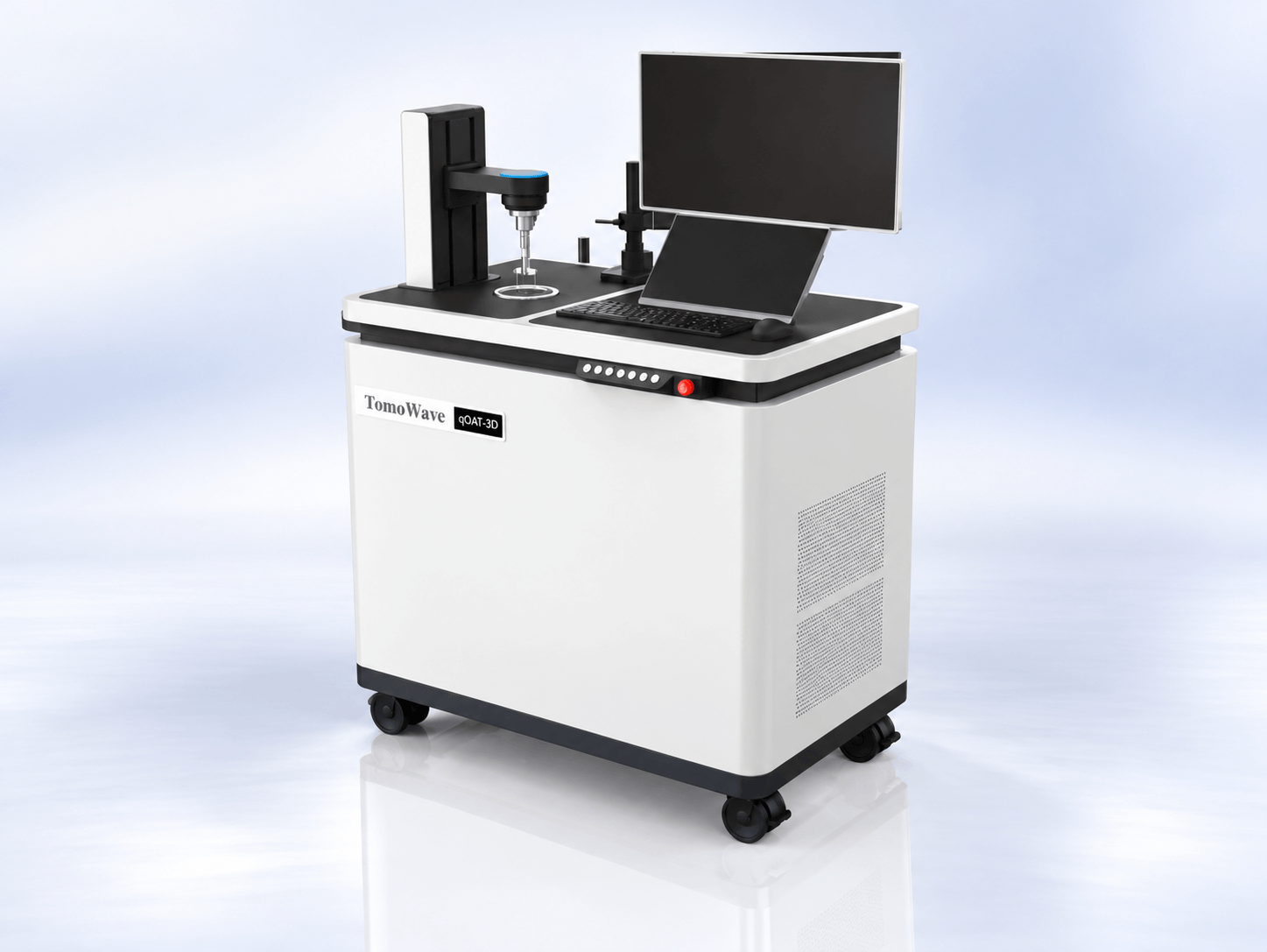
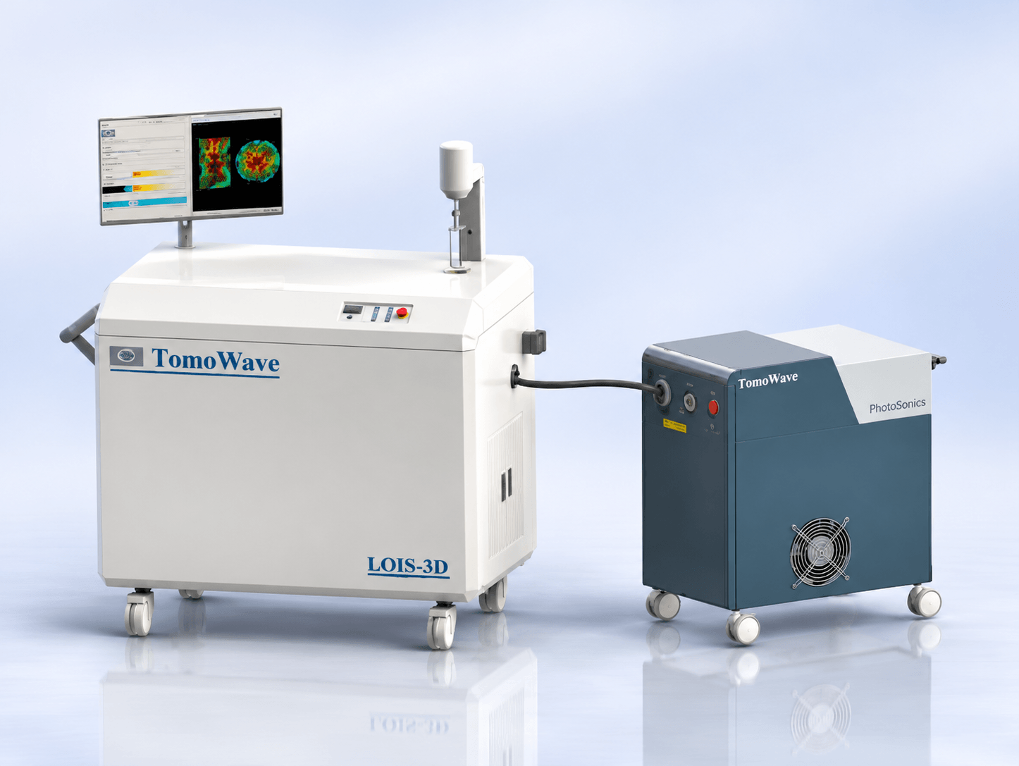
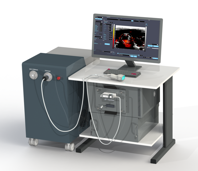
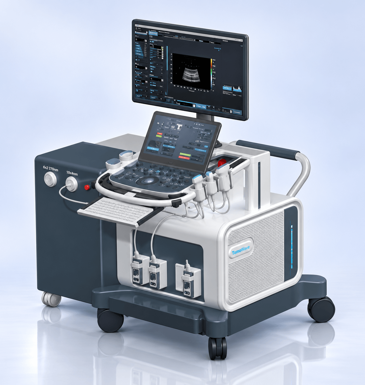
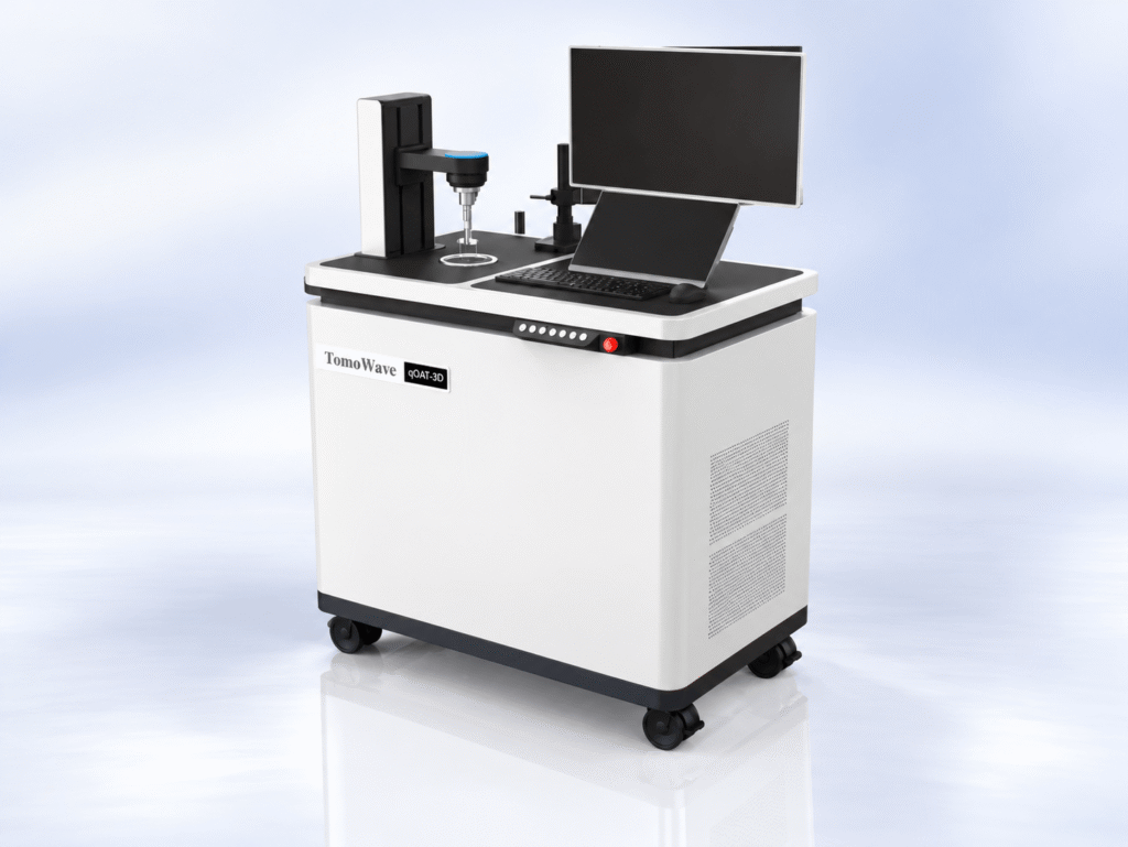
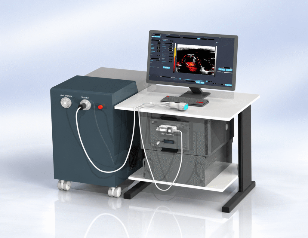
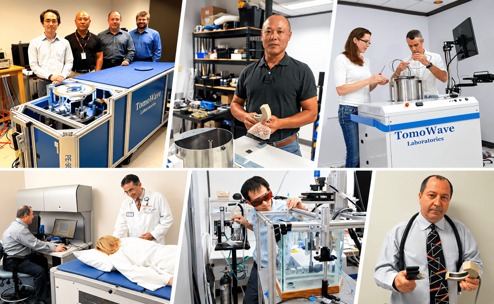
TomoWave develops quantitative optoacoustic tomography systems and superfast high-resolution ultrasound for combined functional, molecular, and anatomical imaging.
Research and Development: from preclinical research to the patient bedside
Imaging for Preclinical Research
Explore high-resolution 3D optoacoustic imaging and ultrasound technology for biomedical research.
Functional and Molecular Imaging
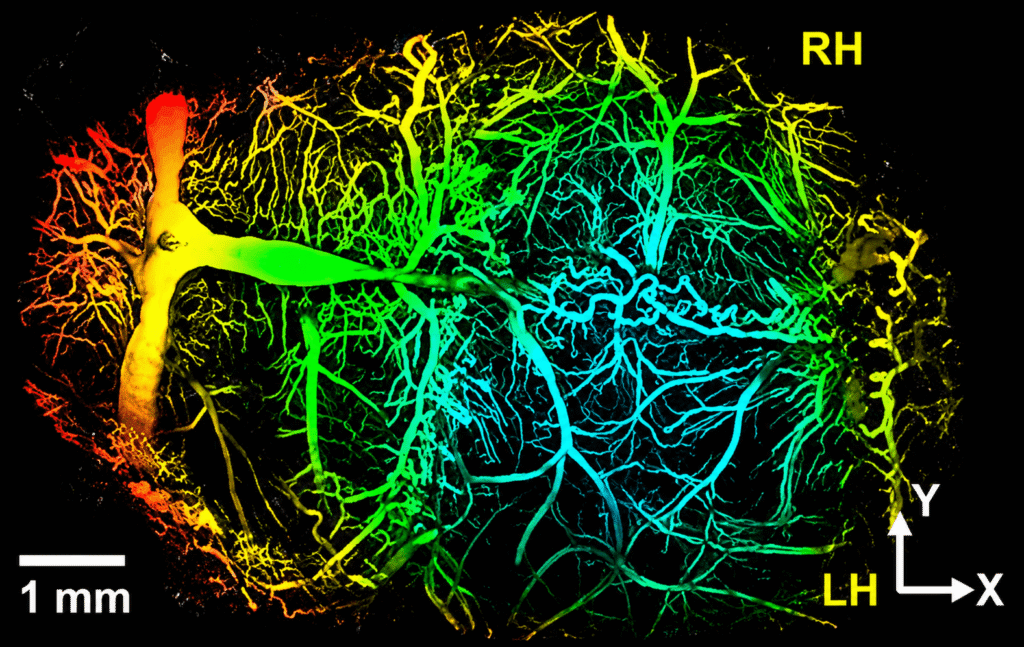
Visualize blood vessels, microvasculature, and tissue oxygenation to study biological functions.
Anatomical Imaging
TomoWave combines the most compelling properties of light and sound to construct detailed images of
biological tissue.
Research and Clinical Applications
TomoWave develops imaging systems for early detection of cancer, vascular disease and image-guided focused ultrasound therapy.
Our Technology
Combining the Properties of Light and Sound
How Optoacoustic Imaging Works
Optoacoustic imaging uses short pulses of laser light and detects resulting ultrasound signals to create detailed molecular images of biological tissues. It helps researchers study tissue structure and functions that may not be visible through conventional imaging alone.
Light Enters the Tissue
Short pulses of near-infrared laser light enter the tissue being studied.
Tissue Molecules Absorb Light
Selected molecules absorb the light energy and produce a very small temperature change.
Ultrasound Signals Are Created
The small temperature change produces pressure waves that travel through the tissue.
Sensors Detect the Signals
Special sensors identify these ultrasound waves and convert them into digital signals.
Detailed Images Are Created
A computer processes the signals to create images that help researchers and doctors study tissue structure with molecular details, blood vessels, and tissue oxygenation.
OUR Technology
Imaging Disease with Light
TomaWave provides MOLECULAR IMAGING with high spatial resolution in the depth of human tissue.
TomoWave develops advanced imaging systems that combine optoacoustic tomography with ultrasound. Our technology helps researchers study both the structure and function of biological tissues
Visualization of vasculature and microvasculature with the goal to detect peripheral vascular disease Early.
Breast Cancer Screening
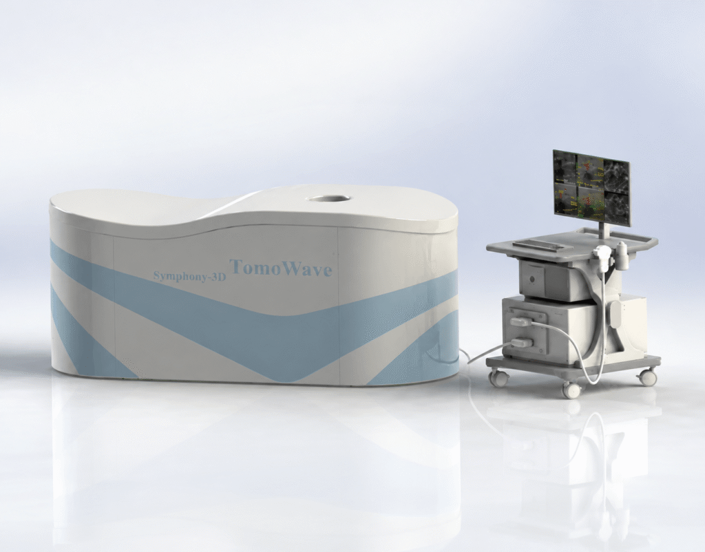
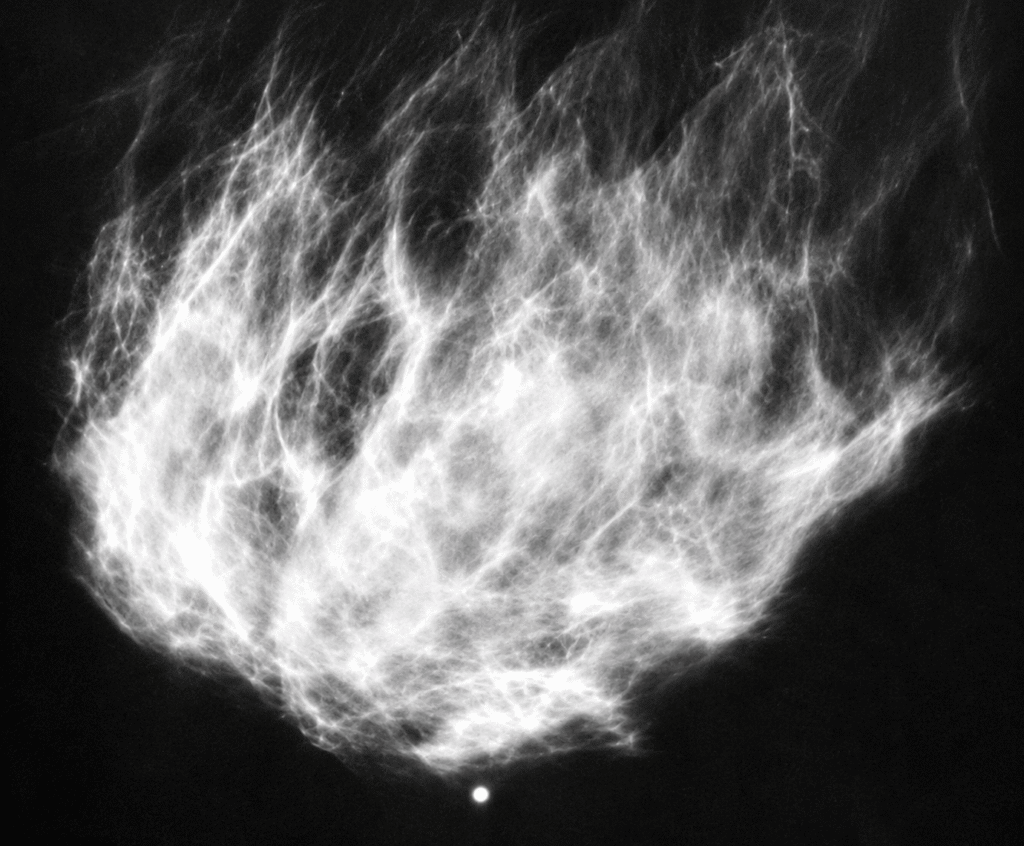
Automated Breast Ultrasound System Enhanced with optoacoustic functional imaging for differentiation of aggressive cancer and benign lesions.
Future developments
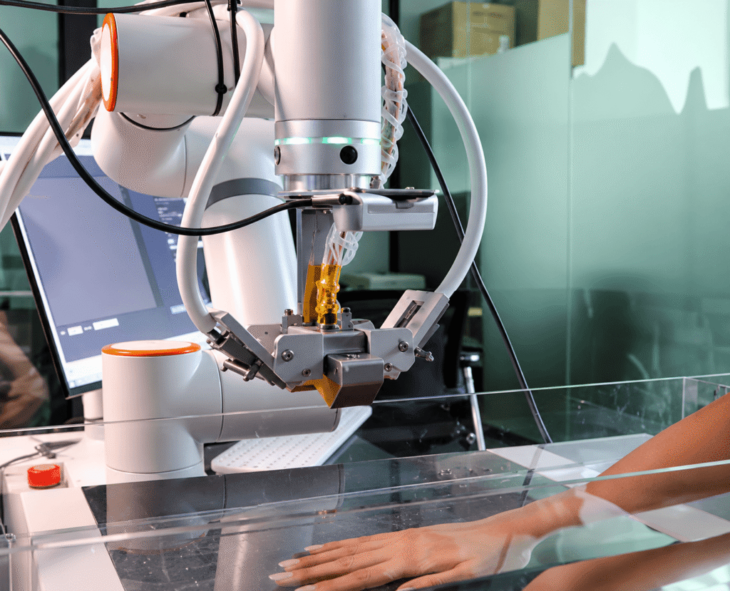
Image-guided focused ultrasound therapy with anatomical visualization provided by ultrasound imaging and functional molecular visualization provided by optoacoustic imaging.
Our Collaborators
See More with TomoWave
Explore how optoacoustic tomography and ultrasound can support your biomedical imaging research.
Discover our technology, get more information about our imaging systems, or contact our team to discuss your research needs.