Our Products

Imaging systems that visualize molecules, their functions and tissue anatomy

Quantitative optoacoustic tomography and ultrasound
for high-resolution, functional, molecular and anatomical imaging.

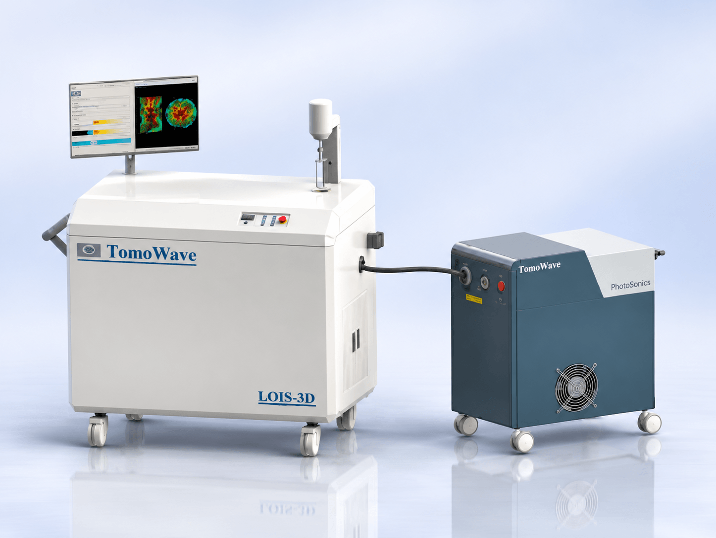
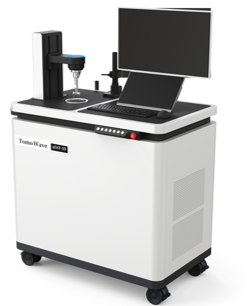
Laser Optoacoustic Imaging System Designed for Biomedical Research in Small Animal Models

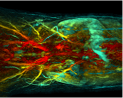
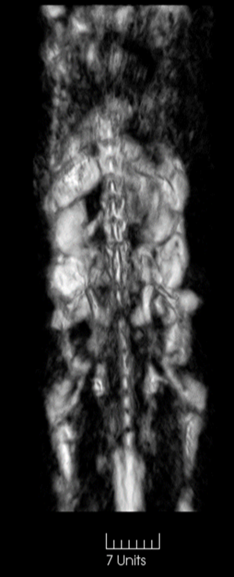
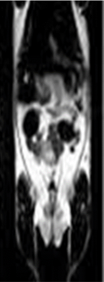
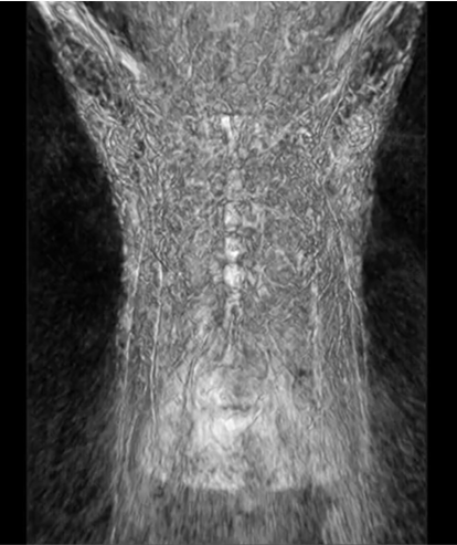
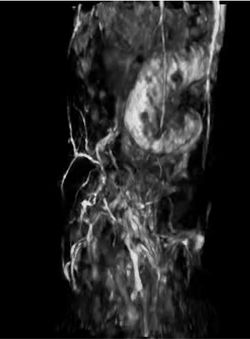
TomoWave Optoacoustic Images

TomoWave Image

MRI Image

TomoWave 3D optoacoustic imaging system has unique advantages in in vivo imaging of small animals due to its high resolution and sensitivity. Combined with the small molecule photoacoustic probes developed by our research group, we have successfully achieved real-time imaging and dynamic distribution of brain signaling molecules.

Zhiyong Jiang, PhD, Professor, Nanjing University, PRC

Products

qOAT-3D

3D quantitative Optoacoustic Tomography System

3D Full view OptoAcoustic Tomography provides the most quantitatively accurate molecular images

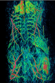
3D OAT Video Image of mouse body organs

3D OAT Video Image of mouse brain

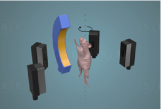
Principles of Full View 3D tomography with 150 micron resolution equal in all 3D

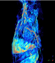
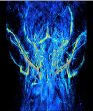
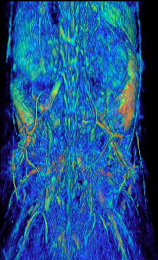
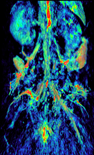
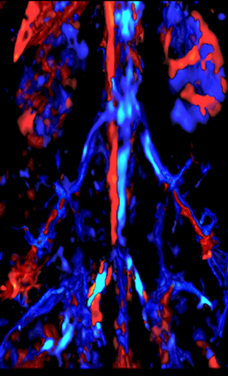
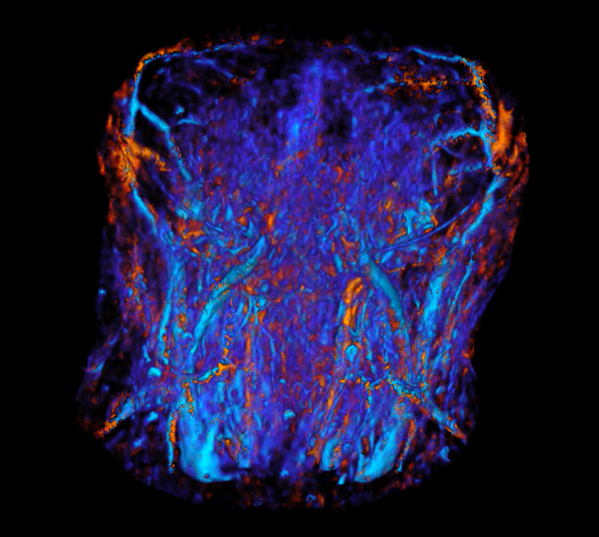
Quantitative 3D functional image of [tHb] and [SO2] through the entire depth of the mouse body

Total Hemoglobin [tHb] 3D Image

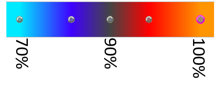
Blood Oxygen Saturation [sO2] Image

Colors presented as a function of brightness

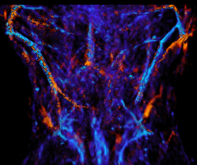
Quantitative 3D functional image of [tHb] and [SO2] through the entire depth of the mouse brain

Products : Products: MAESTRO-RD, Research Imaging System with hand-held probes

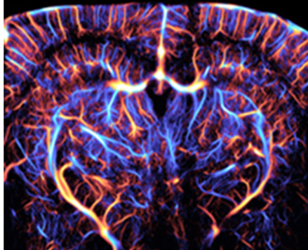
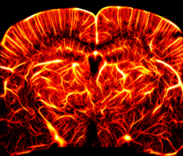
In vivo ULM image – courtesy of Prof. Yachao Zhang, SIBET

MAESTRO-MD: Universal Premium High-End Dual Modality of Ultrasound combined and coregistered with optoacoustics

Future Clinical Applications:

1. Screening diagnostics of early-stage diseases:

  • a. Vasculature and microvasculature in the diabetic foot

  • b. Assessment of kidney’s functional viability prior and after the transplantation

  • c. Functional-anatomical diagnostics of cancer in the breast, brain, thyroid and other organs

2. Differentiation of peripheral arteries, veins, and nerves.

3. Measurements of blood oxygenation in the vital organs.

  • a. Monitoring placenta oxygenation during pregnancy

  • b. Oxygenation of the brain and other various organs

  • c. Detection and diagnostics of sentinel lymph nodes

4. Image-guided surgery: cancer, spine, vasculature, cosmetic.

6. Image-guided focused ultrasound therapy with optoacoustic temperature monitoring.
Microbubbles ULM

Label Free

Transcranial images of a  live mouse brain

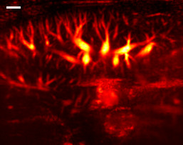
Mouse Liver

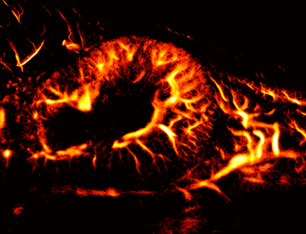
Mouse Kidney

In vivo images – courtesy of Prof. Yachao Zhang, SIBET

Products

SYMPHONY-3D

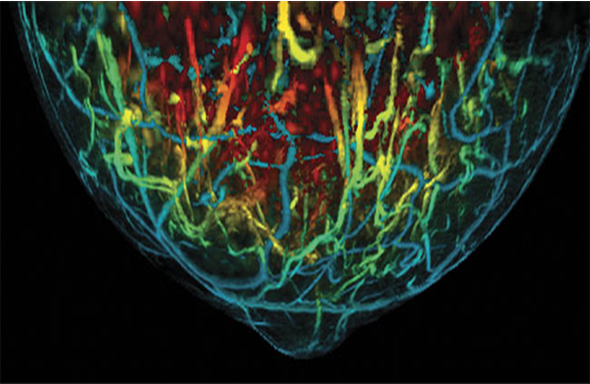
Quantitative Optoacoustic Tomography for Diagnostic Imaging of Breast cancer

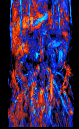
LOUISA-3D OA image of breast vasculature colored vs depth

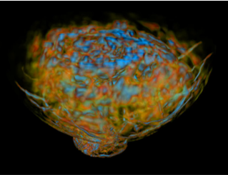
LOUISA-3D OA image of a normal breast

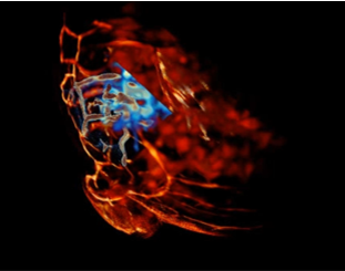
LOUISA-3D OA image of breast cancer defined by hypoxia

Symphony: 3D Automated Screening System for breast cancer

3D Automated Screening System for breast cancer with simultaneous diagnostic capability, not limited by women’s age, breast density and heterogeneity – newly designed functional + anatomical breast scanner. With the scan time potentially down to 5 sec, it will compete with DCE MRI as a safe, more effective and less expensive breast screening modality